Good Monday morning!
On to today's dentistry and health headlines:
Dental Radiographs and Risk for Meningioma
Radiation Risk From Dental Radiography
What Should the Dentist Do?
Dentist charged with lewd conduct
Enjoy your morning!
On to today's dentistry and health headlines:
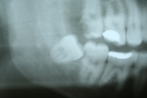
Dental Radiographs and Risk for Meningioma
A recent article by Claus and colleagues[1] reporting an association between dental radiographs and meningiomas has led to renewed concern about the safety of dental x-rays. We will first consider this reported association between dental radiograph and meningiomas, then explore the risks from dental imaging, and finally discuss appropriate use of dental radiographs.
These researchers identified 2228 eligible patients with intracranial meningioma and recruited 2604 matched controls. Participation was 65% for meningioma patients and 52% for controls. Subjects were asked to report their history of bitewing, full-mouth, or panoramic radiographic examinations. The association of meningioma odds with the odds of dental radiographs was calculated by conditional logistic regression for maximum-likelihood estimation of odds ratio, adjusted for age, sex, race, education, and history of head CT.
The researchers reported an association between intracranial meningioma and bitewing radiographs obtained at 10-19 and 20-49 years of age, as well as for all ages combined. Additionally, an association between meningioma and panoramic radiographs at younger than 10 and 10-19 years of age is identified. Finally, an association between meningioma and frequency of panoramic radiographs across all groups, for those with yearly or more frequent radiographs is described. Of note, there was no significant association between intracranial meningioma and full-mouth series. The manuscript concludes that "exposure to some dental x-rays performed in the past, when radiation exposure was greater than in the current era, appears to be associated with an increased risk of intracranial meningioma."
This manuscript, studying the largest cohort of subjects to date, is a significant contribution to existing literature reporting a potential association of intracranial meningioma and dental radiographs.[2-7] However, methodologic details are difficult to evaluate, thus creating ambiguities when performing detailed analysis of the data. For example, although controls were matched for state of residence, local differences do not appear to have been considered. Thus, controls with significantly different dental awareness or access to dental care could have been recruited. No distinction is made between bitewing and selected periapical radiographs. No adjustment for income between patients and controls was performed. The ability of patients and controls to accurately recall history of dental radiographs in a way that would not influence the study findings was not assessed. Finally, the prevalence of dental disease that might affect need for dental radiographs was not evaluated.
Notwithstanding such shortcomings, we believe that a weak association between intracranial meningiomas and dental radiographs may exist.[8] However, current understanding of radiation biology and radiation-induced tumorigenesis (see Radiation Risk From Dental Radiography section below) suggests that an additional 0.02-0.07 mGy to the brain from dental bitewing and panoramic radiography is highly unlikely to contribute measurable risk. Other explanations for such a risk should be considered. An obvious possibility is that patients with intracranial meningiomas have increased odds to receive bitewing or panoramic radiographic examinations.
Radiation Risk From Dental Radiography
The amount of radiation received from dental radiography is so low that it is highly unlikely that it results in a measurable risk. Dose reconstructions using techniques commonly used during the last decades of the last century show that the exposure to the brain from 4 bitewings is approximately 0.07 mGy, and from a panoramic examination about 0.02 mGy. A full-mouth examination (typically consisting of 12 periapical and 4 bitewing exposures) results in a brain dose of approximately 0.24 mGy.
However, even though the brain exposure from a full-mouth examination is higher than from bitewings or a panoramic, the study from Claus and colleagues found associations with the 2 low-dose examinations and meningiomas but not the relatively high-dose full-mouth examination. The resolution of this inconsistency is not clear if radiation is causing the meningiomas.
Further, there are no research reports that support the premise that doses as low as those received by the brain from dental radiography, including from a full-mouth examination, are sufficient to cause meningiomas. We know that brain exposure from dental imaging is much smaller than from head CT examinations. Brain exposure from head CT examinations is typically in the range of 43-75 mGy, far more than from dental radiography.[22] Head CT exposures contribute 4.3% of the collective effective dose from all diagnostic sources, 15 times more than from dental radiography.[23]
What Should the Dentist Do?
Dental radiographs provide a very useful tool in the dentist’s diagnostic armamentarium. Although radiograph benefits outweigh radiation risk,[27] a reasonable and prudent dentist should be cognizant of such a risk.[28,29] It is the dentist's responsibility to consider carefully and justify every radiograph[26] and to employ the means and procedures to optimize radiographic imaging to gain maximum diagnostic information with the minimum radiation.[30]
- Avoid preset intervals for radiographs
- Choose necessary radiographs carefully
- Minimize radiation exposure
Dentist charged with lewd conduct
A Johnson County dentist is charged for doing something lewd while driving.
The woman wants people to know about a man she said exposed himself to her in her Olathe, KS, neighborhood. The man she's talking about is a dentist. Now he's charged with lewd and lascivious conduct, and it was later found out he had been fired from his job after news of the charges.
"On Tuesday night my 3-year-old son and I were out for a jog," the woman, who wished to not have her identity revealed, said.
The woman said she was in an Olathe neighborhood near West 161st Street and South Sunset.
"I saw a silver Honda circling us several times. I thought the guy was lost. Pretty standard in a confusing neighborhood like this," the woman said.
Circling around her, she said was Dr. Wylie Bell.
"I was three houses down from mine, the guy pulls over to the curb, honks his horn," the woman said.
She said what happened next was inexcusable. Bell, a dentist at the time with Arbor Creek Dentistry in Olathe, allegedly exposed himself. When he saw that she wasn't happy about what had just happened, Bell drove off.
"He peeled off and I was able to get his plates, I called the cops and they tracked him down about 45 minutes later," the woman said.
Now Bell is charged with lewd and lascivious conduct, a misdemeanor.
When a KCTV5 crew went to the address listed for Bell, a silver Honda matching the woman's description was parked outside.
"And I learned that he was a professional man. This isn't something you'd expect in a community like this. This isn't something you'd expect with a profession where you're trusting him with your children. It's disgusting," the woman said.
Enjoy your morning!
No comments:
Post a Comment